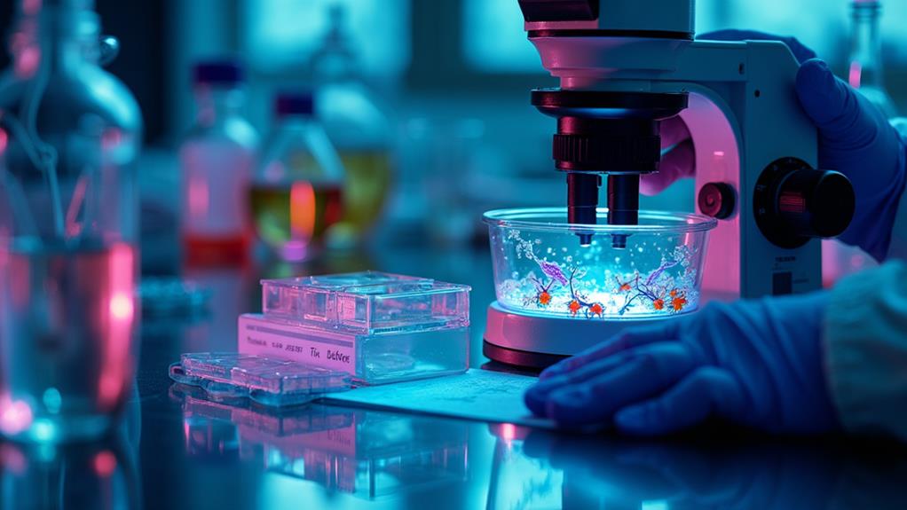
Nanoplankton analysis methods involve a range of sophisticated techniques. You'll find microscopy at the forefront, including light, electron, and fluorescence approaches. Flow cytometry allows for rapid cell counting and characterization, while molecular sequencing provides genetic insights. Imaging flow cytometry combines high-throughput analysis with detailed imaging. Remote sensing applications track nanoplankton dynamics on a global scale, and automated image analysis accelerates data processing. These methods help scientists understand nanoplankton's role in marine ecosystems, carbon cycling, and climate change. Exploring these techniques further will reveal the intricate world of these tiny yet essential organisms.
Microscopy Techniques
Microscopes are the cornerstone of nanoplankton analysis. When you're studying these tiny organisms, you'll primarily rely on light microscopy and electron microscopy techniques.
Light microscopy, including brightfield and phase contrast methods, allows you to observe live specimens and their basic morphology. However, it's limited by the resolving power of visible light.
For more detailed examination, you'll turn to electron microscopy. Scanning electron microscopy (SEM) provides high-resolution images of nanoplankton surfaces, while transmission electron microscopy (TEM) allows you to study their internal structures. You'll need to prepare samples carefully for these techniques, often involving fixation, dehydration, and coating processes.
Fluorescence microscopy is another valuable tool. You can use fluorescent dyes to highlight specific cellular components or exploit the natural autofluorescence of chlorophyll to identify photosynthetic nanoplankton.
Flow cytometry, though not strictly microscopy, complements these methods by allowing rapid analysis of large numbers of individual cells.
When you're working with nanoplankton, it's essential to take into account sample preservation. You'll often need to use fixatives to maintain cell structure, but be aware that these can alter some cellular characteristics.
Flow Cytometry
Flow cytometry steps up the game when it comes to analyzing nanoplankton populations. This powerful technique allows you to rapidly count and characterize individual cells as they pass through a laser beam. You'll find it particularly useful for studying the smallest plankton, which are often difficult to observe with traditional microscopy.
When using flow cytometry, you'll suspend your nanoplankton sample in a fluid stream. As cells pass through the laser, they scatter light and emit fluorescence. You can measure these signals to determine cell size, shape, and pigment content. This method's high-throughput nature means you can analyze thousands of cells per second, providing a thorough view of your sample's composition.
You'll often use fluorescent dyes to label specific cellular components, enhancing the information you can gather. For example, you might stain DNA to differentiate between prokaryotic and eukaryotic cells.
Flow cytometry also allows you to sort cells based on their characteristics, enabling further analysis or culturing of specific subpopulations. This technique's versatility and efficiency make it an indispensable tool in modern nanoplankton research.
Molecular Sequencing Approaches
In recent years, molecular sequencing approaches have revolutionized nanoplankton analysis. These techniques allow you to identify and characterize nanoplankton species at a genetic level, providing unprecedented insights into their diversity and ecological roles.
You'll find that DNA barcoding is a common method, where you sequence specific genetic markers to identify species. For nanoplankton, the 18S rRNA gene is often used. Metabarcoding takes this further, allowing you to analyze entire communities simultaneously.
Next-generation sequencing technologies have greatly enhanced these approaches. You can now generate millions of sequences in a single run, enabling high-throughput analysis of complex nanoplankton assemblages.
Environmental DNA (eDNA) sequencing is another powerful tool. It lets you detect nanoplankton species from water samples without the need for isolation or culturing.
When using these methods, it's essential to evaluate biases in DNA extraction, PCR amplification, and sequencing. You'll also need robust bioinformatics pipelines to process and interpret the large datasets generated.
While molecular approaches offer great advantages, they're best used in combination with traditional morphological and physiological analyses for a thorough understanding of nanoplankton communities.
Imaging Flow Cytometry
Imaging flow cytometry has emerged as a cutting-edge tool for nanoplankton analysis. This technique combines the high-throughput capabilities of traditional flow cytometry with the detailed imaging of microscopy. You'll find it particularly useful for studying nanoplankton, as it allows you to capture individual cell images while simultaneously measuring multiple cellular parameters.
With imaging flow cytometry, you can analyze thousands of nanoplankton cells per second, obtaining both quantitative data and visual information. You'll be able to distinguish between different species based on their morphological features, size, and fluorescence properties. The system captures high-resolution images of each cell, enabling you to perform detailed analyses of cell structure, chlorophyll content, and other cellular components.
You can also use this method to study the physiological state of nanoplankton cells, including their viability and metabolic activity. By incorporating fluorescent dyes or probes, you'll be able to assess specific cellular processes or detect particular molecules of interest.
This powerful combination of imaging and flow cytometry provides you with an all-encompassing tool for characterizing nanoplankton populations in various aquatic environments.
Remote Sensing Applications
Remote sensing applications have revolutionized the study of nanoplankton on a global scale. You'll find that satellite-based sensors can detect ocean color changes caused by nanoplankton blooms, providing valuable data on their distribution and abundance. These sensors measure the spectral reflectance of the ocean surface, which varies based on the concentration and composition of phytoplankton, including nanoplankton.
You can use multispectral and hyperspectral imaging to identify specific nanoplankton species based on their unique optical signatures. This allows you to map their distribution across vast ocean areas.
Additionally, you'll benefit from remote sensing data to track environmental factors that influence nanoplankton growth, such as sea surface temperature, nutrient levels, and light availability.
When you combine remote sensing with in situ measurements and modeling techniques, you'll gain a thorough understanding of nanoplankton dynamics. This integration helps you predict bloom occurrences, assess their impacts on marine ecosystems, and study their role in global carbon cycling.
Automated Image Analysis
Automated image analysis frequently plays an essential role in nanoplankton research. You'll find that this technology allows for rapid processing of large datasets, considerably reducing the time and effort required for manual counting and identification. It's particularly useful when you're dealing with high-throughput sampling methods or extensive field studies.
When you're using automated image analysis, you'll typically employ specialized software that can detect and classify nanoplankton based on their morphological features. These systems often use machine learning algorithms to improve accuracy over time. You'll need to train the software with a diverse set of labeled images to guarantee it can recognize various species and morphotypes.
One of the key advantages you'll notice is the consistency in measurements and classifications. Unlike manual analysis, which can be subject to human error and bias, automated systems provide reproducible results.
However, you should be aware that these systems aren't infallible. You'll still need to perform quality checks and occasionally verify results manually, especially for rare or unusual specimens.
As you integrate automated image analysis into your workflow, you'll likely see considerable improvements in the efficiency and scale of your nanoplankton research.

Erzsebet Frey (Eli Frey) is an ecologist and online entrepreneur with a Master of Science in Ecology from the University of Belgrade. Originally from Serbia, she has lived in Sri Lanka since 2017. Eli has worked internationally in countries like Oman, Brazil, Germany, and Sri Lanka. In 2018, she expanded into SEO and blogging, completing courses from UC Davis and Edinburgh. Eli has founded multiple websites focused on biology, ecology, environmental science, sustainable and simple living, and outdoor activities. She enjoys creating nature and simple living videos on YouTube and participates in speleology, diving, and hiking.
🌿 Explore the Wild Side!
Discover eBooks, guides, templates and stylish wildlife-themed T-shirts, notebooks, scrunchies, bandanas, and tote bags. Perfect for nature lovers and wildlife enthusiasts!
Visit My Shop →
